Introduction
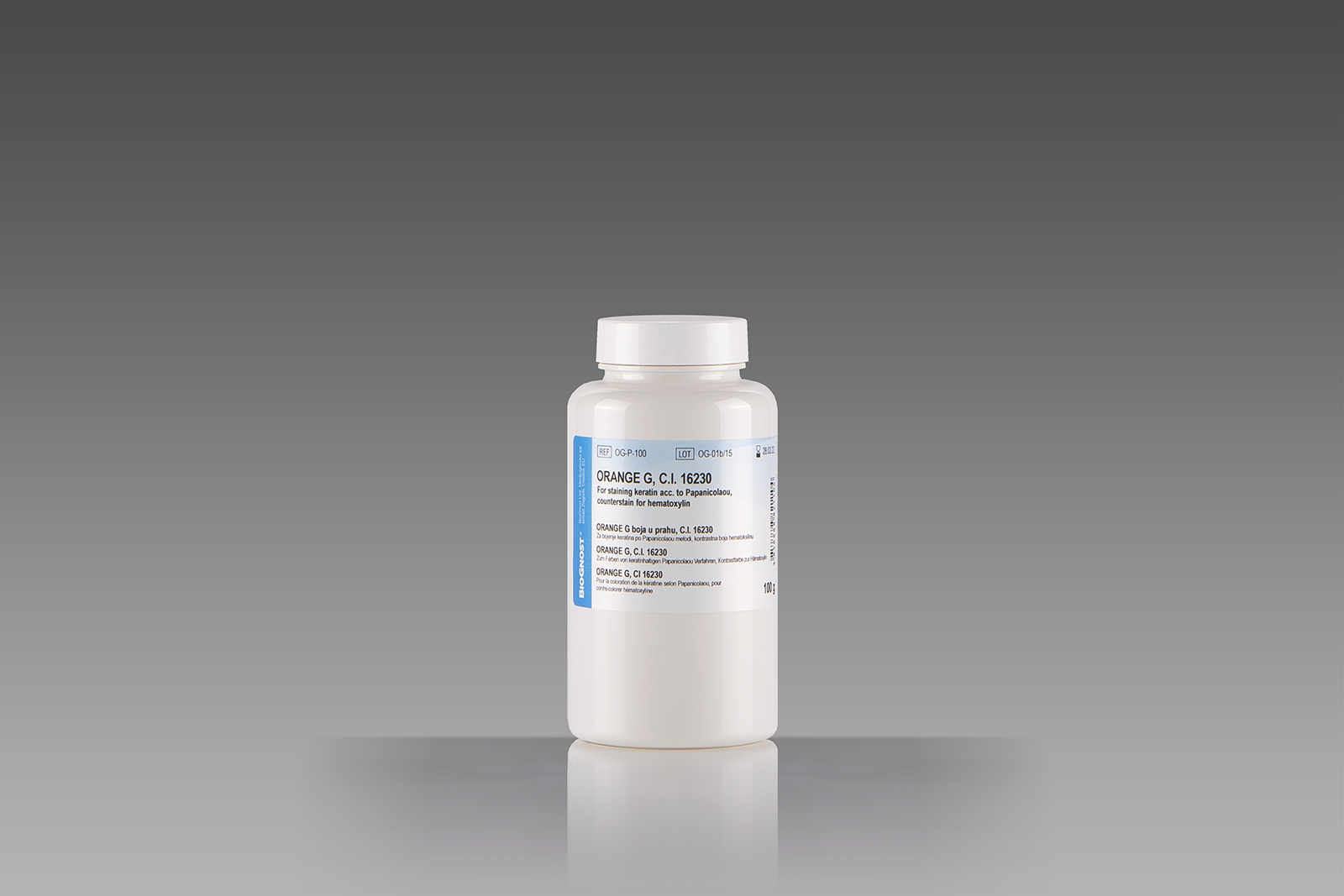
Histology, cytology and other related scientific disciplines study the microscopic anatomy of tissues and cells. In order to demonstrate a good tissue and cellular structure, the samples need to be stained in a correct manner. Orange G powder dye is used in various staining methods in microscopy. It is a component of many biological dyes, usually as a background dye or cytoplasmic dye, and the most commonly used method of staining cytological samples is according to Papanicolaou. During the first step of staining, nuclei are being stained blue, dark purple or black using hematoxylin. The second step consists of cytoplasmic staining using an orange solution, and targeted structures are stained orange in various intensities. During the third step the so-called polychromatic solution is used – a mixture of eosin, Light Green S.F. and Bismarck Brown dye. In histopathology, Orange G is a part of the Mallory connective tissue dye.


