Introduction
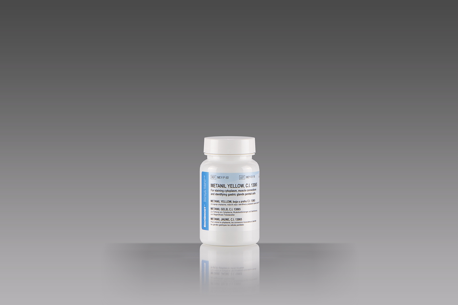
Histology, cytology and other related scientific disciplines study the microscopic anatomy of tissues and cells. In order to achieve a good tissue and cellular structure, the samples need to be stained in a correct manner. Metanil yellow, small-molecule dye, acts as a light-lipophilic anion in aqueous solutions (except in strongly acid solutions). Metanil yellow is most commonly used for counterstaining in Mucicarmine staining method (acc. to Mayer or in various modifications), but also for cytoplasmic staining in different polychromatic procedures, such as method acc. to Masson (developed by Lillie), that is, for cytoplasmic counterstaining (Gridley’s visualization of fungi in histopathology sections; method acc. to Clark). Combined with P.A.S. reagent, Metanil yellow is used for visualization of ciliary muscle connectors (Schachar). Metanil yellow-P.A.S.-Toluidine Blue staining enables the identification of parietal cells in gastric glands and in gastric epithelium.


