Introduction
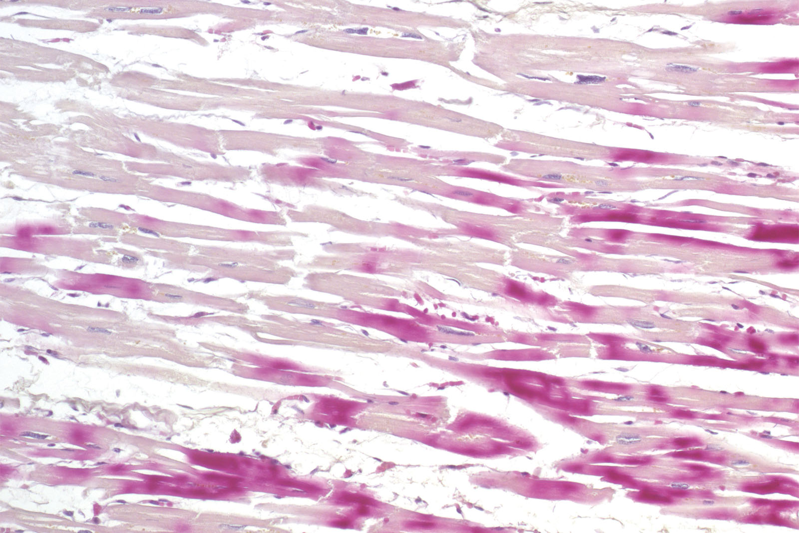
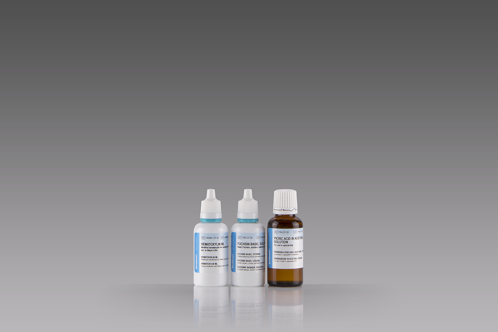
Histological diagnosis of ischemia in the early phase of myocardial infarction using the standard hematoxylin-eosin histological methods and a light microscope is exceptionally delicate. The reason for that is minimal histopathological changes occurring on the cardiac muscle during the first six hours of symptoms. However, staining the section using the kit consisting of hematoxylin, basic fuchsin and picric acid enables a histological overview of early changes on the cardiac muscle caused by ischemia or myocardial infarction.


