Introduction
An impeccable sample fixation is a prerequisite for a correct histological diagnosis. Tissue samples must be immersed in an optimally chosen fixative immediately after sampling because a timely fixation will prevent autolysis, putrefaction and other unwanted cellular changes. Although there are hundreds of histological fixatives and at least tens of formaldehyde-based fixatives, neutral buffered formaldehyde solutions with a concentration range from 4% to 10% are the most commonly used fixatives, primarily because of their simple and universal application. Tissue fixation using a buffered formaldehyde solution results in forming cross-links, i. e. it forms methylene bridges between proteins, that is, it results in keeping tissue components in their in vivo relation. If fixated properly, the tissue sample can withstand additional histological tissue processing and staining.
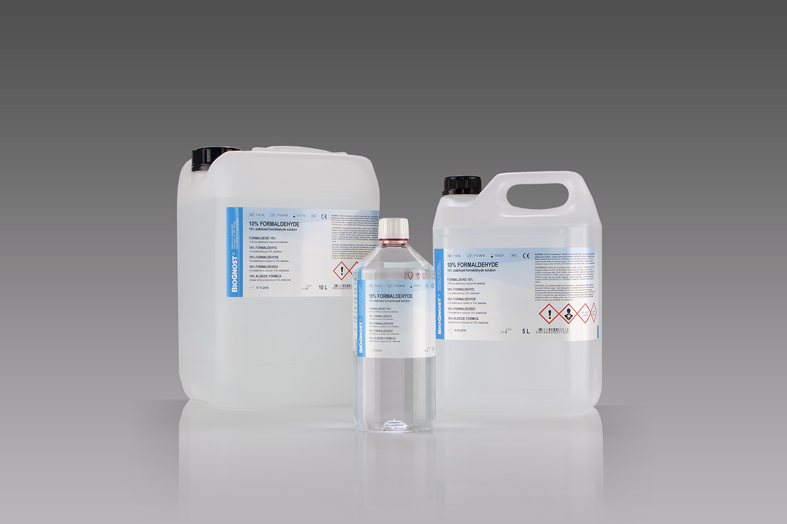
10% formaldehyde solution is a 25% formalin, which is the most commonly used fixative. Suitable for fixing larger tissue samples. It is a solution ready for use, colorless and with a characteristic odor, suitable for fixating larger tissue samples. It is a primary tissue fixative. Tissue samples can be fixated through a shorter period of time and with a lower volume ratio towards fixated tissue in comparison to 4% formaldehyde solution. Added methanol prevents formaldehyde polymerization. It is suitable for usage in all automated devices for tissue processing, as well as for manual histological techniques. It is conveniently packaged in 1 liter bottles, 5, 10 and 20 liter canisters


