Introduction
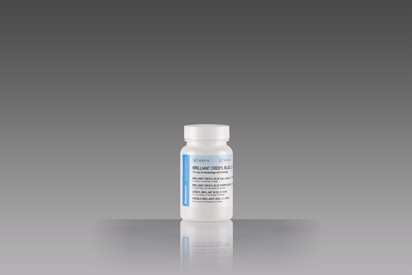
Supravital staining is a method of staining used in microscopy to examine living cells that have been removed from an organism. It differs from intravital staining, which is done by injecting or otherwise introducing the stain into the body. Thus a supravital stain may have greater toxicity, as only a few cells need to survive it for a short period of time. As the cells are alive and unfixed, outside the body, supravital stains are temporary in nature. The most common supravital stain is performed on reticulocytes using brilliant cresyl blue, which makes it possible to see the reticulofilamentous pattern of ribosomes characteristically precipitated in these live immature red blood cells by the supravital stains. By counting the number of such cells the rate of red blood cell formation can be determined, providing an insight into bone marrow activity and anemia.


