Special staining kits for histology and citology
-
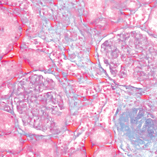
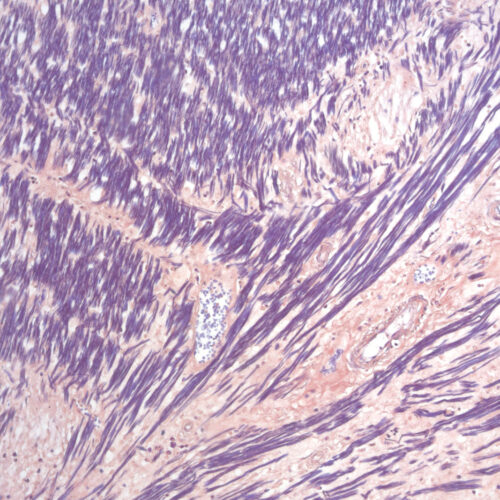
Gomori Trichrome kit
Five-reagent kit for staining muscle, collagen fiber and nuclei, contains blue counterstain. The kit can be used to contrast skeletal, cardiac or smooth muscle.
-
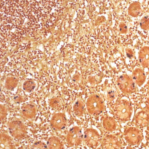
Grimelius kit
Five-reagent kit for staining argyrophilic granules. Grimelius kit can be used for the detection of secretory intracytoplasmatic granules specific for carcinoid tumors and for identification of neuroendocrine cells.
-
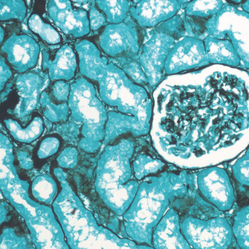
Grocott kit, stabilized
Seven-reagent kit for visualization of fungi and histological argentaffin structures in general (such as basal membranes). Green counterstain provides clear and visually rich contrast to target structures stained black.
-
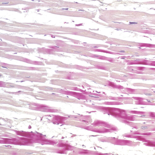
H.B.F.P. kit
Three-reagent Hematoxylin-Basic Fuchsin-Picric acid staining kit for detection of cardiac muscle changes after ischemia or myocardial infarction. H.B.F.P. kit is a non-enzymatic histochemical technique for detection of early myocardial ischemia with vivid contrast.
-
Hematoxylin P.T.A. kit
Four-reagent Hematoxylin-Phosphotungstic Acid kit for differentiation of smooth and striated muscle tissues as well as for the detection of fibrin, collagen and elements of the central nervous system according to Mallory.
-
Hematoxylin W kit
Acid-resistant hematoxylin according to Weigert. Two-reagent kit that stains the nuclei blue-black, often a component of special staining kits for connective tissues.
-
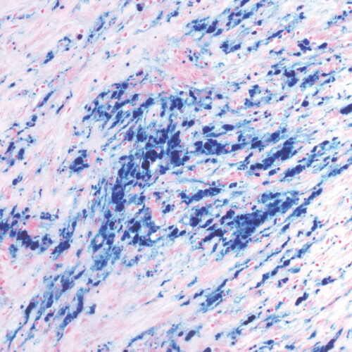
Hemognost Perls kit
Three-reagent HemoGnost Perls (Prussian blue / Berlin blue) kit for the detection of reactive ferric (Fe3+) ions in cells. It is often applied on bone marrow and spleen cells.
-
Ladewig’s Trichrome kit
Four-reagent kit for staining muscle (brown to red) and collagen fibers with blue counterstain. Staining procedure is easier and faster compared to Masson and Mallory Trichrome staining procedures.


