Histology and cytology
-
UriGnost SM reagent
Modification according to Sternheimer-Malbin for staining and microscopical analysis of urine sediment. A component of UriGnost SM kit.
-
Van Gieson Trichrome kit
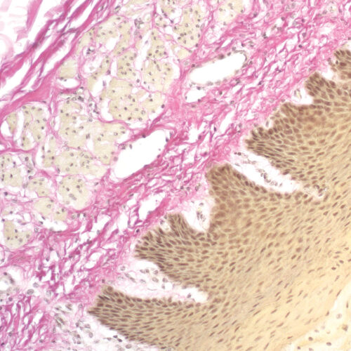
Three-reagent kit for staining collagen fibers, muscle tissue, keratinized epithelium, cytoplasm, glial fibers and erythrocytes. Used for differentiation between collagen and smooth fibers in tumors and various other diseases.
-
Verhoeff kit
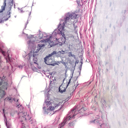
Six-reagent kit for detecting atrophy of elastic tissue in cases of emphysema, thinning and loss of elastic fibers in arteriosclerosis and other vascular diseases, or whether blood vessels have been invaded by a tumor.
-
Victoria Blue R, C.I. 44040
Basic Blue 11, Victoria Lake Blue R. For use in histology, cytology and microbiology.
-
Von Kossa kit
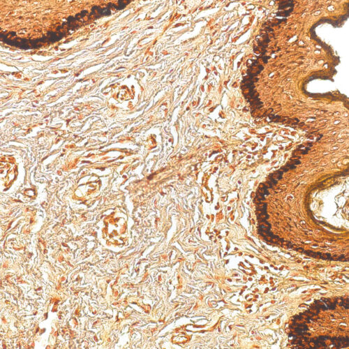
Five-reagent kit for simple and reproducible detection of calcium deposits and calcium salt in tissue samples according to Von Kossa. Tissue calcification is associated with metabolic problems in various tissues (like bone marrow and mamma) and in tumors.
-
Warthin Starry kit
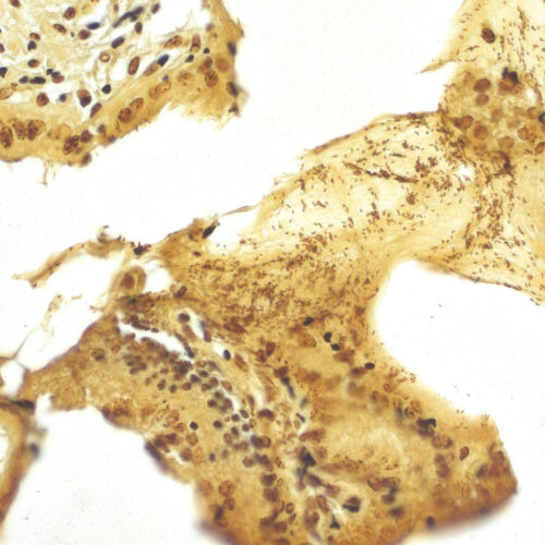
Five-reagent kit for staining Spirochaeta, Helicobacter pylori, Microsporidia and Legionella pneumophila. The kit contains 12 jars with gelatin that enables both incubation and staining of sections, as well as other reagents that enable precipitation of silver on the bacterial surface. The bacteria are found in the mucus of the surface epithelium, in the apical gastric glands and in the gastric mucosa.
-
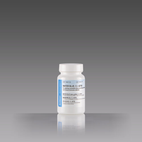
Water Blue, C.I. 42755
Acid Blue 22, BSC certified stain. For staining connective tissue according to Mallory and Gomori trichrome methods.


