Histology and cytology
-

-
-
-
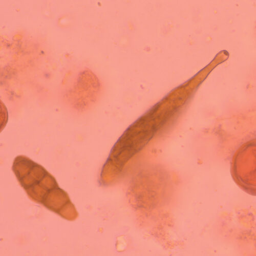
M.I.F. kit
Two-reagent merthiolate-iodine-formalin kit for fixing and staining fecal parasites (especially protozoa, cysts, helminthic eggs and larvae). The stool sample is fixed with formalin and stained with two coloring agents; iodine and Eosin Y.
-
Malachite Green, C.I. 42000
Malachite Green Oxalate, Basic Green 4, Victoria Green, BSC certified stain. Used as counterstain in TB-Stain Cold kit.
-
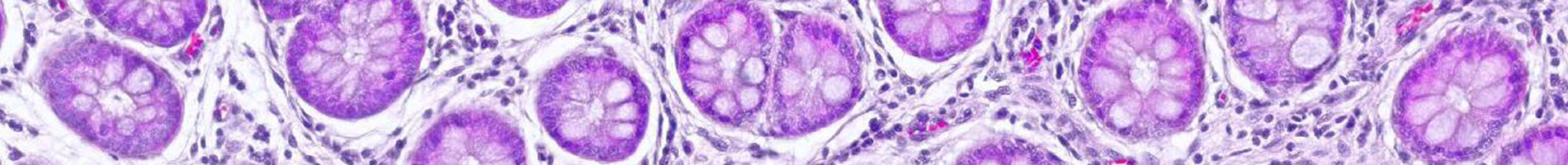
Mallory Trichrome kit
Three-reagent staining kit for connective tissue visualization and detection of collagen, cartilage, muscle, elastic fibers, mucous, pituarity cells, reticulum, bones, amyloid and erythrocytes.
-
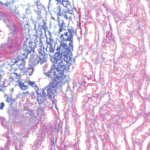
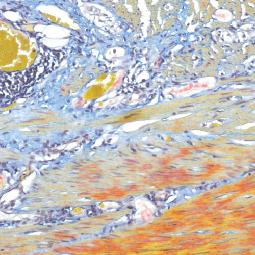
Martius Scarlet Blue (MSB) kit
Seven-reagent kit used for fibrin visualization, especially of older clusters. This method is a modification of Masson Trichrome and is ideal for studying connective tissue and vascular pathology.
-
Martius Yellow C.I. 10315
Golden Yellow, Martinsgelb, Acid yellow 24. For staining erythrocytes yellow in trichrome staining methods.